When radiation therapy successfully targets a brain tumour, it feels like a victory – and it is. But for a subset of patients, that victory comes with a delayed and deeply unsettling consequence: radiation necrosis, the progressive death of healthy brain tissue in the very region that was treated.
Managing this condition has historically posed a clinical dilemma. Steroids can suppress inflammation temporarily; surgery carries its own risks; and not every patient is a suitable candidate for invasive intervention. This is precisely where Hyperbaric Oxygen Therapy (HBOT) is rewriting the narrative.
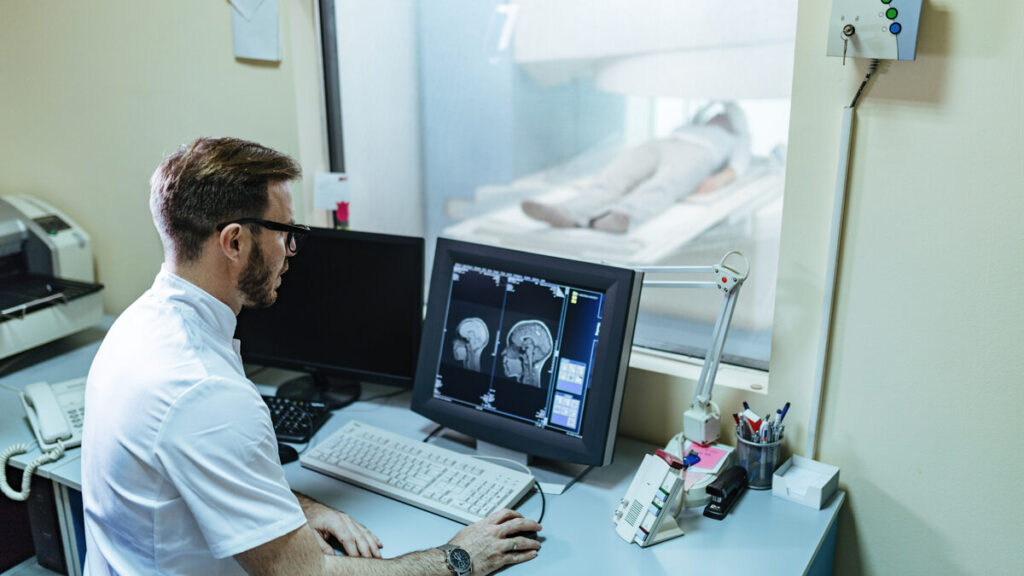
By delivering 100% pure oxygen at elevated atmospheric pressure, HBOT addresses radiation necrosis at the biological root – stimulating angiogenesis, reversing ischemic tissue damage, and restoring perfusion to oxygen-starved brain tissue. At Vayu Prana, HBOT Center in Kolkata, we’ve seen firsthand how this therapy can offer measurable, lasting relief even when other options fall short. Here’s a comprehensive breakdown of the science, the evidence, and what patients can realistically expect.
What Is Hyperbaric Oxygen Therapy?
The name itself is instructive: hyper (increased) + baric (pressure) = therapy delivered under elevated pressure conditions. In practice, HBOT involves breathing 100% pure oxygen inside a pressurised chamber at approximately two to three times normal atmospheric pressure.
Under these conditions, oxygen dissolves directly into the blood plasma – not just into haemoglobin – significantly increasing the total oxygen concentration available to tissues throughout the body. This makes HBOT uniquely valuable in situations where oxygen delivery is compromised and blood transfusion is either unavailable, contraindicated, or ruled out for immunological or religious reasons.
Radiation necrosis is one such situation – and increasingly, HBOT is being recognised not just as a supplementary option, but as a frontline non-surgical intervention.
What Is Radiation Necrosis?
Radiation necrosis is a late-onset complication of radiotherapy or radiosurgery targeting Central Nervous System (CNS) tumours or metastases. It occurs when high-dose radiation – particularly doses exceeding 55 Gray (Gy) – damages the surrounding brain parenchyma beyond its capacity to repair itself.
The timeline is deceptive. Most patients develop symptoms within a year of completing radiotherapy, but the condition can surface as late as six to seven years post-treatment. Its presentation on imaging frequently mimics tumour recurrence, making accurate diagnosis genuinely challenging.
How common is it?
A 2007 study found that approximately 2.8% of patients treated for malignant glioma developed focal radiation necrosis. Among those who survived beyond one year post-treatment, that figure climbed to 9%.
Definitive diagnosis typically requires one of two approaches: sustained radiological monitoring through serial scans and imaging, or histopathological confirmation through surgical resection and biopsy.
What Causes Radiation Necrosis?
At the cellular level, radiation necrosis is driven by vascular injury – damage to the fine network of blood vessels supplying brain tissue.
Here’s the biological cascade:
- Radiation induces vascular injury → damaged vessel walls begin leaking plasma fluid into surrounding tissue (cerebral oedema).
- Oedema triggers VEGF release → Vascular Endothelial Growth Factor (VEGF), previously known as Vascular Permeability Factor, is released in response to the injury. VEGF disrupts the blood-brain barrier (BBB), causing further capillary leakage.
- BBB breakdown worsens oedema → The cycle becomes self-reinforcing. As oedema builds, brain parenchyma becomes increasingly hypoxic and susceptible to necrotic change.
This is not an acute injury – it is a slow, progressive deterioration that can unfold over months or years after radiation exposure has ended.
Risk Factors for Radiation Necrosis
A 2016 study identified several patient- and treatment-specific factors that significantly influence the likelihood of developing radiation necrosis:
- Lesion location within the brain
- Lesion size at the time of treatment
- Radiation dose – 55 Gy is considered the upper safe threshold
- Older age – ageing reduces vascular resilience and capillary integrity
- Diabetes – pre-existing vascular compromise accelerates injury
Understanding these risk factors allows clinicians to identify high-risk patients early and implement proactive monitoring protocols.
How Is Radiation Necrosis Diagnosed?
Radiation necrosis typically presents within the first year after radiotherapy ends. Patients may report one or more of the following:
- Persistent or severe headaches
- Focal neurological deficits (weakness, speech difficulty, vision changes)
- Drowsiness or unusual fatigue
- Personality or behavioural changes
- Cognitive impairment or memory disturbances
- Confusion or disorientation
- Seizures in more advanced cases
Because these symptoms closely resemble tumour recurrence, distinguishing radiation necrosis from relapse requires sustained radiological follow-up using MRI and advanced imaging modalities – or, when imaging is inconclusive, surgical biopsy.
Treatment Options for Radiation Necrosis
All treatment strategies for radiation necrosis share the same foundational goal: reducing cerebral oedema to protect surviving brain parenchyma from further damage. Current options include:
Corticosteroids (e.g., Dexamethasone) with Vitamin E
The first-line pharmacological approach. Effective in reducing inflammatory oedema, but long-term steroid use carries its own risks, including immunosuppression and metabolic effects.
Bevacizumab (Anti-VEGF Antibody)
Directly neutralises VEGF – the key driver of abnormal vascular permeability – thereby interrupting the oedema cascade at its source. Bevacizumab has shown meaningful radiological and clinical improvement in several trials.
Surgical Resection
Reserved for severe or rapidly deteriorating cases. Carries inherent operative risks, particularly in eloquent brain areas or when necrosis is multifocal.
Laser Interstitial Thermal Therapy (LITT)
A minimally invasive ablative technique suited for accessible lesions, though not universally available or applicable.
Hyperbaric Oxygen Therapy (HBOT)
The preferred non-surgical option for patients who are poor operative candidates, have lesions in surgically inaccessible locations, or present with multiple necrotic sites. Increasingly supported by clinical evidence as a standalone intervention.
The Role of HBOT in Treating Radiation Necrosis
Radiation necrosis is, at its core, an ischaemic and coagulative process – tissue dies because it is starved of oxygen. HBOT addresses this directly, and through multiple complementary mechanisms:
1. Reversing Hypoxia
By flooding tissues with 100% oxygen at elevated pressure, HBOT restores oxygen availability to ischaemic regions of brain parenchyma – the precise mechanism needed to interrupt the necrotic process.
2. Reducing Cerebral Oedema
HBOT-mediated vasoconstriction helps reduce capillary leak and cerebral oedema, directly counteracting the vascular permeability that drives tissue damage. The National Cancer Institute has recognised HBOT as an adjunct treatment for radiation-induced oedema, particularly when used alongside Dexamethasone.
3. Stimulating Angiogenesis
HBOT mobilises bone marrow-derived stem cells, which differentiate into endothelial cells capable of forming new blood vessels. This neo-angiogenesis creates fresh vascular pathways into damaged tissue, increasing perfusion across the disrupted blood-brain barrier.
4. Promoting Collagen Synthesis and Vessel Repair
Beyond new vessel formation, HBOT promotes collagen production that supports structural repair of damaged capillaries – ultimately restoring capillary density to approximately 80% of pre-injury levels.
Clinical Evidence: What Trials Show
2020 Adjunct Study
A 2020 clinical study evaluated HBOT as an adjunct to corticosteroid therapy in patients with brain radiation necrosis. The results demonstrated significant clinical stabilisation and measurable radiological improvement across multiple patients, with a notable additional benefit: steroid dosage requirements decreased both during and following the course of HBOT.
Case Study: HBOT as a Standalone Treatment
Perhaps the most compelling evidence for HBOT’s independent efficacy comes from a single-patient case study that offers an unusually clear before-and-after picture.
Patient profile: A 43-year-old male presented 18 months after total surgical resection and adjuvant radiotherapy for a right temporal astrocytoma. Pre-treatment MRI revealed an area of abnormal white radiopacity in the right temporal lobe extending across the midline. The patient reported headaches rated 8/10 on the pain severity scale.
Treatment choice: After being offered steroids, bevacizumab, or HBOT, the patient selected HBOT exclusively.
Protocol: Each session comprised three 30-minute intervals of 100% oxygen delivered at 2 atmospheres of pressure in a hyperbaric chamber.
Outcomes:
- After 32 sessions: MRI showed a visible reduction in radiopacity intensity; headache severity dropped by approximately 50%.
- After 60 sessions: MRI demonstrated near-complete resolution of the necrotic lesion with significantly reduced oedema. The patient reported that headache frequency and intensity had declined to near zero within two months.
This case stands as robust evidence that HBOT – administered without any concurrent pharmacological intervention – can independently arrest and reverse the progression of radiation necrosis.
Safety Profile of HBOT
HBOT is among the safest available therapeutic modalities when administered within established clinical parameters. Key safety considerations include:
Session structure matters: Limiting sessions to under two hours and maintaining pressures below 3 atmospheres significantly reduces the risk of adverse effects. The three-part, 30-minute session format used in radiation necrosis protocols is well within this safe window.
Common mild effects: Patients may experience ear or sinus discomfort (due to pressure changes), temporary light-headedness, or post-session fatigue. These are generally transient and self-resolving.
Oxygen toxicity: In rare cases, prolonged or excessive oxygen exposure can trigger seizures. Research indicates this risk is concentrated in patients with specific cofactors – including narcotic or alcohol withdrawal, or concurrent use of antidepressants, cephalosporins, ceftriaxone, or tramadol. Pre-treatment screening for these risk factors is a non-negotiable part of responsible HBOT practice.
Dose individualisation: Oxygen dosage and session frequency should always be determined on a per-patient basis by a qualified clinician, with periodic breathing of normal air incorporated into longer protocols to prevent oxygen accumulation.
Conclusion
Radiation necrosis is a serious complication – but it is not untreatable. For patients facing this diagnosis, Hyperbaric Oxygen Therapy offers something genuinely rare in this clinical context: a non-surgical, biologically targeted intervention with a compelling evidence base and a strong safety profile.
What makes HBOT particularly compelling is its versatility. It can function as an adjunct to corticosteroid therapy, as an alternative to bevacizumab, or – as the case evidence suggests – as a standalone treatment capable of producing near-complete lesion resolution. For patients who are not candidates for surgery, or whose necrosis is multifocal or anatomically inaccessible, HBOT may represent the single most viable path forward.
That said, every patient’s situation is unique. The decision to pursue HBOT should be made in close consultation with a qualified clinician who can assess individual risk factors, review imaging, and design a protocol tailored to the patient’s specific condition and medical history.
At HBOT Center in Kolkata, our approach is precisely that – personalised, evidence-informed, and always focused on what is safest and most effective for you.
Frequently Asked Questions (FAQs)
Q1: How many HBOT sessions are typically required to treat radiation necrosis?
The number of sessions varies based on lesion severity, location, and individual patient response. Clinical evidence suggests meaningful improvement can begin within 30–40 sessions, with near-complete resolution achieved in some patients after 60 sessions. Your treating clinician will define a protocol specific to your case.
Q2: Can HBOT be used alongside other radiation necrosis treatments?
Yes. HBOT is well-documented as an effective adjunct to corticosteroid therapy, and it can complement bevacizumab treatment. In fact, combining HBOT with Dexamethasone has been shown to allow a reduction in steroid dosage while maintaining or improving clinical outcomes.
Q3: Is HBOT painful or uncomfortable?
HBOT itself is not painful. Patients lie inside a pressurised chamber and breathe normally. Some individuals experience mild ear or sinus discomfort during pressurisation – similar to the sensation during an aircraft descent – which is easily manageable with slow pressure adjustments.
Q4: Who is not a suitable candidate for HBOT in radiation necrosis?
Patients with certain medical risk factors require careful evaluation before commencing HBOT. These include individuals in narcotic or alcohol withdrawal, or those currently taking antidepressants, ceftriaxone, cephalosporins, or tramadol, due to elevated seizure risk. A thorough pre-treatment medical review is essential.
Q5: How long after radiotherapy can radiation necrosis develop, and when should HBOT be considered?
Radiation necrosis most commonly presents within one year of completing radiotherapy, but it can emerge up to six or seven years later. HBOT should be considered at diagnosis, particularly for patients who are poor surgical candidates, have multifocal necrosis, or prefer a non-pharmacological approach.
Q6: Is HBOT available in India for radiation necrosis treatment?
Yes. HBOT Center in Kolkata provides hyperbaric oxygen therapy for radiation necrosis and other conditions, with individualised treatment protocols developed in consultation with experienced clinicians. Early referral following radiotherapy for CNS tumours is encouraged for at-risk patients.