Dermal fillers have transformed modern aesthetics – offering a non-surgical path to smoother skin, fuller lips, and more defined facial contours. But like every medical procedure, they carry risks that can escalate quickly without timely, informed intervention. Among the most feared complications is vascular occlusion – a condition where filler material enters or compresses a blood vessel, cutting off the vital supply of oxygen and nutrients to surrounding tissue. When left untreated, it can rapidly progress to tissue necrosis and permanent scarring.
When conventional first-line treatments fall short, Hyperbaric Oxygen Therapy (HBOT) has emerged as a powerful, evidence-backed solution. By delivering 100% pure oxygen inside a pressurized chamber, HBOT floods oxygen-deprived tissues with the supply they urgently need – even when normal blood flow is compromised. The result: reduced swelling, accelerated wound repair, and a significantly lower risk of irreversible tissue damage.
In this guide, we break down exactly how vascular occlusion develops after filler injections, what warning signs to watch for, and why HBOT is increasingly becoming the treatment of choice for managing this complication – backed by clinical evidence and expert insight from HBOT India.
Understanding Dermal Fillers
What Are Dermal Fillers and How Do They Work?
Dermal fillers are injectable substances used to restore lost facial volume, smooth deep wrinkles, and enhance features such as lips and cheekbones. As we age, the natural collagen framework beneath our skin breaks down – causing the skin to thin, sag, and lose its youthful plumpness. Fillers counteract this by physically replenishing lost volume where it matters most.
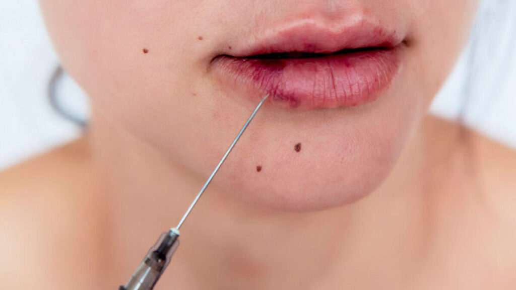
The procedure is straightforward: a trained practitioner marks the treatment area and applies a topical anesthetic to minimize discomfort. Filler is then introduced in small, precise amounts using a fine needle. Following the injections, an ice pack is typically applied to reduce swelling and discomfort. Most patients resume normal activity immediately.
While results can be dramatic and gratifying, the procedure is not without risk. Beyond the common side effects of bruising and swelling, more serious complications – including infection, vascular occlusion, and tissue necrosis – can arise, particularly when the filler inadvertently interacts with the vascular system.
Dermal Fillers and Vascular Occlusion
1. What Is Vascular Occlusion?
Vascular occlusion occurs when filler material enters the bloodstream or compresses a blood vessel from the outside, restricting or completely blocking the delivery of oxygen and nutrients to surrounding tissues. It can present as either partial occlusion – where blood flow is reduced – or complete occlusion, where blood flow is entirely cut off.
At its mildest, vascular occlusion causes redness (erythema) and bruising. At its most severe, it can result in ischemia and, if not promptly addressed, full tissue necrosis – permanent, irreversible cell death.
2. How Do Dermal Fillers Cause Vascular Occlusion?
Vascular occlusion during filler procedures can occur through three primary mechanisms:
- Intravascular embolism – filler is accidentally injected directly into a blood vessel and travels downstream, lodging in a smaller vessel and blocking blood flow.
- Extra-vascular compression – the filler mass, once deposited, physically presses against a nearby vessel from the outside.
- Vascular spasm – the injection triggers involuntary constriction of the vessel wall.
Once filler enters a blood vessel, it may migrate toward progressively smaller vessels – those with limited backup (collateral) circulation – where it causes localized ischemia. In high-pressure injection scenarios, the bolus can even travel in a retrograde direction, moving against normal blood flow and reaching distant vessel branches. This explains why ischemic symptoms can appear far from the original injection site.
Additionally, intramuscular injection can inadvertently trigger local clotting mechanisms (thrombolytic activity), adding another pathway to vessel blockage. In every scenario, the priority of treatment is the same: remove the injected material as quickly and completely as possible.
3. Delayed Vascular Occlusion
Not all occlusions present immediately after injection. Some develop hours later due to:
- Hygroscopic fillers (such as certain hyaluronic acid products) that absorb water over time, gradually expanding and exerting pressure on adjacent vessels.
- An embolus lodging in a vessel with weak collateral circulation, causing ischemia to develop progressively as nutrient deprivation accumulates.
- Filler delivered near – but not inside – a vessel (infra-arterial injection) that initially appears fine but later triggers platelet aggregation and progressive blockage.
- Injections near bifurcation points that lead to delayed occlusion in terminal vessel branches.
Signs and Symptoms of Vascular Occlusion
Early recognition is critical. The following signs warrant immediate medical attention:
- Blanching: Sudden whitening of the skin due to interrupted blood supply. This is usually the first visible indicator of occlusion. Note: transient blanching caused by lidocaine or vasoconstrictors during the procedure is harmless and resolves quickly – true occlusion-related blanching does not self-resolve.
- Bruising: Discoloration ranging from blue to dull purple-gray, indicating vessel damage or blockage in the area.
- Swelling: While some swelling is a normal post-filler side effect, abnormal or worsening swelling – particularly when accompanied by other symptoms – may indicate vascular compromise.
- Skin darkening or blackening: Progressive discoloration is a serious warning sign of tissue necrosis. Once necrosis takes hold, the damage is irreversible.
- Unusual or escalating pain: Pain that intensifies after the procedure – particularly beyond the injection site – can indicate occlusion. Local anesthetic may initially mask this symptom.
- Skin coolness: A noticeable drop in skin temperature at or near the injection site is a strong indicator of reduced blood circulation.
Conventional Treatment of Vascular Occlusion
When vascular occlusion is detected early, a number of standard interventions may be employed:
- Massage, warming, and tapping of the affected area to encourage blood flow.
- Hyaluronidase injections – the enzyme used to dissolve hyaluronic acid-based fillers and reverse occlusion in cases where the filler is HA-based.
- Antibiotic therapy to address or prevent infection stemming from necrotic tissue.
- Superficial debridement – removal of dead tissue to facilitate wound healing.
When these measures fail to produce adequate improvement, combination therapy with HBOT becomes the next line of intervention.
Hyperbaric Oxygen Therapy (HBOT) for Vascular Occlusion
What Is HBOT?
Hyperbaric Oxygen Therapy involves breathing 100% pure oxygen inside a sealed, pressurized chamber. The atmospheric pressure within the chamber is elevated above normal sea-level conditions, which dramatically increases the amount of oxygen that dissolves directly into the blood plasma. This allows oxygen to reach tissues even when red blood cell delivery is impaired by vessel blockage — bypassing the very bottleneck that vascular occlusion creates.
HBOT has a well-established history in wound care and plastic surgery, and the Federal Council of Medicine in Brazil formally recognized it for clinical use as far back as 1995. Its safety and efficacy profile has since been validated by numerous medical bodies, including the Brazilian Society of Hyperbaric Medicine.
How HBOT Addresses Vascular Occlusion Specifically
HBOT acts on vascular occlusion through multiple complementary mechanisms:
- Hyper-oxygenation of plasma: Pressurized oxygen dissolves into blood plasma, enabling oxygen delivery to ischemic tissues regardless of vessel blockage.
- Reduction of edema: HBOT has vasoconstriction properties that help reduce fluid accumulation and swelling around the occlusion site.
- Angiogenesis stimulation: HBOT activates stem cells and promotes the formation of new capillaries (angiogenesis), rebuilding microcirculation in damaged tissue.
- Enhanced bactericidal activity: Oxygen-rich environments restore phagocyte function in hypoxic tissues, strengthening the immune response against infection.|
- Fibroblast proliferation: HBOT promotes the activity of fibroblasts, the cells responsible for producing collagen and repairing structural tissue.
- ROS and VEGF activation: Reactive Oxygen Species (ROS) and Vascular Endothelial Growth Factor (VEGF) – both stimulated by HBOT – play critical roles in wound healing and infection control.
Crucially, HBOT must be initiated as early as possible after occlusion is identified. The sooner oxygen-deprived tissues are treated, the greater the chance of halting necrotic progression and promoting meaningful recovery.
Recommended HBOT Protocol
While treatment protocols are individualized based on clinical severity, established guidelines suggest:
- For uncomplicated vascular occlusion: two HBOT sessions per day for the first seven days, followed by one session daily for a further week.
- For cases involving necrotizing fasciitis or significant tissue necrosis: up to 30 sessions over 5–6 weeks, at a pressure of 2–3 ATA (atmospheres absolute) per session.
Therapy continues until clinical improvement is confirmed and necrotic progression has halted.
Clinical Evidence Supporting HBOT for Filler Complications
A peer-reviewed case study published in the Dermatology Journal documented two patients who developed vascular occlusion following hyaluronic acid filler injections. Both presented with skin discoloration and prolonged capillary refill times – hallmark indicators of compromised circulation. Initial treatment with hyaluronidase injections over two days produced no meaningful improvement.
Both patients were subsequently placed on an HBOT protocol: two sessions daily, breathing 100% oxygen at pressurized conditions, for a minimum of seven days. This was followed by one session per day for a further week. By the end of the treatment course, both patients showed clear clinical improvement – demonstrating the value of HBOT as a rescue therapy when first-line treatments are insufficient.
This case, along with growing clinical literature, supports the integration of HBOT into the standard management pathway for moderate-to-severe filler-related vascular complications.
Frequently Asked Questions (FAQs)
1. What is vascular occlusion, and how does it happen after a filler injection?
Vascular occlusion occurs when filler material accidentally enters or compresses a blood vessel, blocking the normal supply of oxygen and nutrients to surrounding tissue. It can result from direct intravascular injection, external compression by the filler, or vascular spasm triggered by the needle. In some cases, the material migrates to smaller vessels – including distant ones – leading to ischemia well beyond the injection site.
2. How quickly does vascular occlusion develop after a filler procedure?
Vascular occlusion can present immediately after injection or be delayed by several hours. Immediate occlusion typically manifests as sudden skin blanching or pallor at the site. Delayed occlusion may appear hours later due to the hydrophilic nature of certain fillers that gradually attract water and compress nearby vessels, or due to progressive platelet accumulation at an injection site that worsens blood flow over time.
3. What are the early warning signs of vascular occlusion I should not ignore?
Key warning signs include sudden skin blanching at or near the injection site, unusual or escalating pain after the procedure, visible bruising or discoloration (blue to gray-purple), localized skin coolness, and swelling that exceeds the normal post-filler response. If the skin begins to darken or turn black, tissue necrosis may already be underway – this is a medical emergency requiring immediate intervention.
4. How does HBOT help treat vascular occlusion from dermal fillers?
HBOT delivers 100% oxygen inside a pressurized chamber, allowing oxygen to dissolve directly into blood plasma at levels far beyond what normal breathing achieves. This mechanism bypasses compromised blood vessels and delivers oxygen to ischemic tissue. HBOT simultaneously reduces edema, stimulates angiogenesis, activates stem cells, and enhances the immune system’s infection-fighting capacity – providing a comprehensive biological response to vascular occlusion damage.
5. How many HBOT sessions are typically needed for filler-related vascular occlusion?
Session protocols vary depending on severity. Published case studies have reported two HBOT sessions daily for the first seven days, followed by one session daily for an additional week. For cases involving tissue necrosis, up to 30 sessions over 5–6 weeks may be recommended. Your treating physician will tailor the protocol based on your clinical presentation and response to therapy.
6. Is HBOT safe for patients who have had dermal filler complications?
Yes. HBOT has a well-established safety profile and has been used in wound care and plastic surgery for decades. It is non-invasive and works alongside – rather than instead of – standard treatments such as hyaluronidase and antibiotics. Most patients tolerate HBOT well. A thorough physician assessment is always recommended before initiating therapy.
7. Can HBOT reverse tissue necrosis caused by vascular occlusion?
HBOT cannot reverse necrosis that has already occurred – established tissue death is generally irreversible. However, HBOT is highly effective at halting the progression of necrosis by oxygenating viable surrounding tissue, reducing local inflammation, and promoting healing at wound margins. Early initiation of HBOT significantly improves the chance of limiting damage and achieving the best possible outcome.
Conclusion On HBOT for Vascular Occlusion
Vascular occlusion following a dermal filler procedure is a time-sensitive medical emergency – every hour without adequate oxygenation increases the risk of permanent tissue damage. While early intervention with hyaluronidase and supportive care remains the critical first step, Hyperbaric Oxygen Therapy offers a uniquely powerful adjunct when conventional approaches are insufficient.
By delivering a high concentration of oxygen directly to ischemic tissues under pressure, HBOT targets the core consequence of vascular occlusion: cellular oxygen starvation. Its ability to reduce edema, stimulate angiogenesis, activate stem cells, and enhance microcirculation makes it one of the most comprehensive tools available for managing filler-related vascular complications.
Clinical case studies continue to validate its use, and institutions like HBOT India are at the forefront of applying this therapy for aesthetic medicine complications. If you or someone you know experiences signs of vascular occlusion after a filler procedure – blanching, unusual pain, or progressive skin discoloration – seek expert medical attention immediately. Early access to HBOT could be the critical difference between full recovery and irreversible damage.